Patients who suffer from severe aortic stenosis now have a minimally invasive procedural option available at South Texas Health System Heart. Transcatheter aortic valve replacement (TAVR) is an advanced heart valve replacement procedure that offers new hope to patients who have aortic valve stenosis and are at moderate or high risk for open-heart surgery.
Aortic Stenosis
Aortic stenosis is a common heart condition where the buildup of calcium deposits causes a narrowing of the aortic valve opening. These deposits prevent the valve from opening and closing properly, which makes the heart work harder to pump blood to the body. This can cause the heart to weaken and function poorly over time, which can lead to heart failure and an increased risk for sudden cardiac death.
Symptoms of aortic stenosis may include:
- Chest pain or pressure
- Dizziness
- Fatigue
- Fainting
- Heart murmur
- Heart palpitations
- Rheumatic fever
- Shortness of breath during activity
Treating Aortic Stenosis
Treatment is typically managed by a team of experienced cardiologists and cardiovascular surgeons who collaborate to determine the most appropriate care for each patient. Medications do not cure aortic stenosis but are sometimes prescribed to help control symptoms, maximize heart function, control blood pressure and control irregular heart rhythm.
Aortic valve replacement (AVR) is an invasive surgical procedure and is the standard treatment for patients with aortic stenosis. Fortunately, for those patients considered high risk or ineligible for surgery, transcatheter aortic valve replacement (TAVR) provides a new option.
How TAVR Works
During the minimally invasive TAVR surgery, the cardiologist replaces the heart valve without open-heart surgery and without removing the diseased valve. TAVR is performed while the heart is beating; therefore, there is no need for a heart-lung machine. A TAVR valve is made of biological material and is supported with a metal stent.
A catheter is inserted through an artery in the leg and then guided through the arteries into the pumping chamber of the heart under direct guidance imaging. The catheter may also be introduced through an artery in the chest or directly through the aorta. The heart valve is compressed and placed on a balloon catheter. The valve is positioned inside the diseased aortic valve. The balloon valve is then inflated to secure the valve in place. This enables the new valve to take over the original valve’s function, allowing oxygen-rich blood to flow efficiently from the heart to the rest of the body.
Meet Our Team
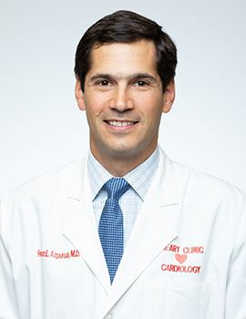
 Federico Azpurua, MD
An interventional cardiologist hailing from the Universidad Central de Venezuela, Dr. Azpurua has been trained at St. Elizabeth’s Medical Center, Texas Heart Institute, Baylor College of Medicine and more. He is fluent in both English and Spanish.
A few of his sub-specialties include:
- Peripheral Vascular Disease and Endovascular Intervention
- Structural Heart Disease
- Echocardiography
- Nuclear Cardiology
 Carlos D. Giraldo, MD, FACC
Hailing from the Temple University School of Medicine, Dr. Giraldo received specialty training from Emory University School of Medicine, The Ochsner Clinic Foundation and The University of Florida—SHANDS.
Some of his specialties include:
- Interventional Cardiology with Vascular/Peripheral training
- Structural Heart Disease
- General Cardiology with special training in Advanced Heart Failure and Cardiac Transplantation
- Nuclear Cardiology
